MLTC-1 mouse testicular interstitial cell tumor cells
BNCC number: 360318
Growth properties : Adherent growth
Growth conditions: 37 ℃,5% CO2
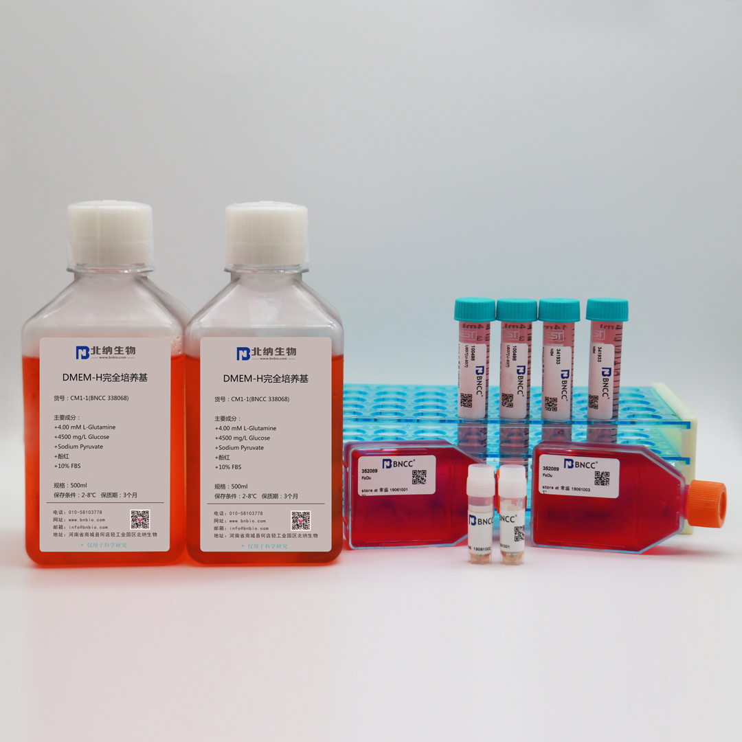
Complete medium: 80% 1640 + 20% FBS
Cryopreservation conditions: 50% basal medium + 40% FBS + 10% DMSO
Receiving notice: if any abnormality is found on the day of receipt, please contact the customer service within 24 hours. If it is overdue, it is deemed that the cells are well. For the frozen cells, it shall be stored in the refrigerator at -80 ℃ upon arrival. If they are not used for a long time, they shall be transferred to liquid nitrogen for storage overnight. Recovered cells in T25 culture flask, upon receipt, put the culture flask in the incubator for 2-3h, and then carry out handling procedure. During recovery, each vial shall be used up once and shall not be retained. After recovery, the cells can be passed on to the next generation and can be used normally. Please operate in strict accordance with this instruction, otherwise the replacement of cells are not be available in case of loss of cell viability.
Recovery steps:
(1)The frozen vial is taken out of liquid nitrogen or -80 ℃ refrigerator and put into PE gloves, quickly submersed into a 37 ℃ water bath, shaken the frozen vial to accelerate dissolution, and it is advisable to dissolve all within 1min;
(2)Put the dissolved cell solution into a centrifuge tube containing 9ml of complete culture medium in an ultra-clean table, centrifuge at 1000-1200rpm for 5min, discard the supernatant, resuspend the cells with 1-2ml of complete media.
(3)The cell suspension was added to T25 flask containing 5-6ml complete medium and cultured in an incubator.
Subculture:
(1)remove the medium, rinse twice with PBS , and add 1-2mL pancreatin (0.25% Trypsin + 0.02% EDTA);
(2)Observe the digestion situation under the microscope. When the cell edge shrinks and the adherent is loose (a pasteur pipette can be used to suck up some pancreatin and gently blow somewhere in the cell layer, and the cell layer can be seen to detach with naked eyes, I .e. digestion is completed, otherwise digestion is continued), directly suck out pancreatin, add 5-6mL of complete medium, gently blow the cell layer off.
(3)dispense the cell suspension into a fresh T25 flask as the ratio of 1:2, add appropriate complete culture medium, make the cell suspension well distributed, and culture in an incubator.
(4)Pay attention to the change of pH value of media and cell density, renew the media regularly (2-3 times a week), and repeat the passage operation or cryopreservation when the cell density reaches 80%-90%.
Notes:
(1)The cells are density dependent. Initiate the subcultivation in T25 flask when the cell density reaches 80%; A subcultivation ratio of 1:2 is recommended.
(2)If the cells in sealed culture bottle, put it into the incubator for cultivation after handling, with the cap unscrewed.
Recovery record: According to the recovery instructions, the results of the cell recovery are reported as follows:
| Item |
quality standard |
recovery record |
| viability: |
adherence is observed in 18 hours, the cell adherence rate ≥ 80.0% in 90hours |
adherence is observed in 18 hours, the cell adherence rate ≥ 80.0% in 80hours |
| cell morphology: |
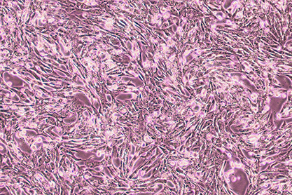
epithelial cell-like |
epithelial cell-like, long strip |
| attached figure: |
 |
 |
| Conclusion: |
good viability, and no abnormal cell morphology, qualified |

 info@bncc.com
 - English
 - Japanese

